
Kneecap Chondromalacia: What It Means and How It’s Treated
Understand kneecap chondromalacia, how it differs from runner’s knee, what scans can show, and why treatment usually starts with rehabilitation.
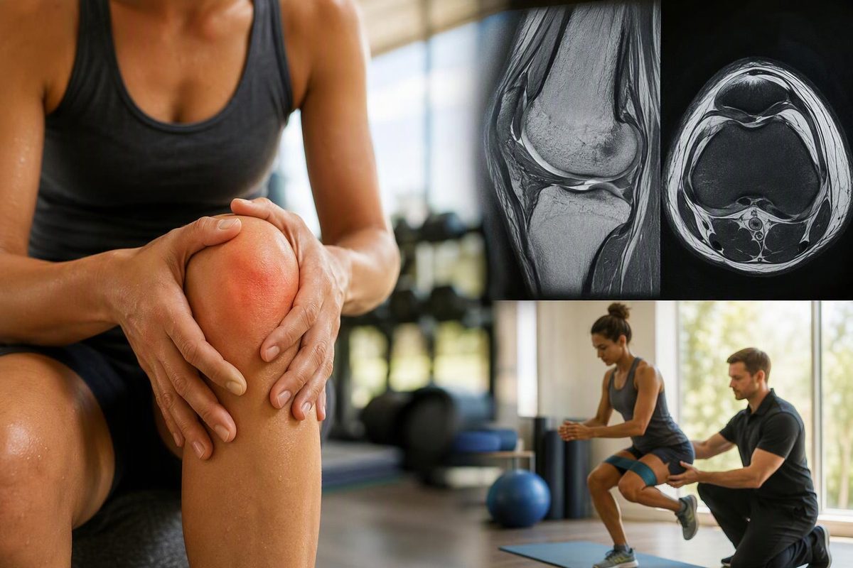
Kneecap chondromalacia means softening or damage of the smooth cartilage on the underside of the kneecap, also called the patella. This cartilage helps the kneecap glide against the thighbone as you bend and straighten your knee. The term describes a tissue change—not something you can diagnose from stair pain or grinding alone. AAOS explains the cartilage changes and their relationship to knee pain.
If the term appears in your clinic letter or MRI report, the useful next question is: does this finding explain my symptoms, and does it change my treatment?
Is chondromalacia the same as runner’s knee?
Not exactly. Patellofemoral pain, often called runner’s knee, describes pain around or behind the kneecap. Chondromalacia describes changes in the cartilage itself. They can occur together, but someone can have patellofemoral pain without demonstrated cartilage damage.
That distinction matters because cartilage has no pain-sensing nerves. Associated pain can come from underlying bone or irritated tissues around the joint, rather than directly from the cartilage. Pain intensity is therefore not a reliable measure of how much cartilage is damaged. AAOS and a condition-specific research review explain the sources of pain and the difficulty relating symptoms to cartilage damage.
Possible symptoms include an ache around or behind the kneecap, pain on stairs or after prolonged sitting with bent knees, and a grinding sensation called crepitus. These overlap with other knee conditions; none confirms chondromalacia by itself. Patient’s clinician-reviewed overview describes these patterns.
Why does the cartilage change?
There may be several contributors rather than one clear cause. Repeated loading, a knee injury, kneecap instability, or the shape and alignment of the kneecap joint can contribute. Cartilage changes can also occur with ageing. It is not simply a condition of runners, nor does the diagnosis prove that your exercise technique caused it. Patient and the research review outline the possible contributors.
How is it diagnosed—and do you need a scan?
Assessment starts with your symptom history and an examination of knee movement, kneecap tracking, strength and stability. A clinician may use a working diagnosis and start treatment without imaging, particularly when the examination suggests uncomplicated patellofemoral pain. That does not mean cartilage damage has been confirmed. Patient explains provisional diagnosis and testing.
When tests are useful:
- X-rays help assess bones, alignment and arthritis-related changes, but are not good at identifying early chondromalacia.
- MRI can show cartilage lesions and other structures inside the knee. It is not perfect, particularly for subtle changes, and its findings need to be interpreted alongside the examination.
- Arthroscopy, a procedure using a camera inside the joint, allows direct inspection. It is invasive and is not a routine next step simply because the knee hurts or grinds.
These strengths and limitations are described in the chondromalacia review. Persistent symptoms or uncertainty about the diagnosis may justify further investigation.
What does a cartilage grade mean?
A report may grade the damage. Grading systems differ, but a common four-grade system runs from softening and swelling at grade 1 to full-thickness cartilage loss exposing underlying bone at grade 4. The grade describes the cartilage, and the severity does not reliably track symptoms. Research review.
Do not read the grade as a pain score, recovery deadline or automatic need for surgery. Ask which system was used and how the finding affects your plan.
Treatment usually starts with rehabilitation
Initial treatment generally focuses on easing symptoms and improving function:
- Adjust aggravating activity. Temporarily reduce strenuous knee loading rather than repeatedly provoking pain. Choose alternative activity according to what your knee tolerates.
- Use an individualised exercise plan. Physiotherapy can build thigh strength and address relevant movement or muscle-balance problems.
- Use symptom relief when appropriate. Pain medicines may help in the short term; ask a pharmacist or clinician what is safe for you.
These are standard conservative approaches described by Patient.
For people with patellofemoral pain, a 2024 best-practice guide recommends education and knee-targeted exercise, with hip exercise and measures such as taping or foot orthoses selected according to individual needs. That evidence supports treating the accompanying pain and functional difficulties; it does not establish that exercise regrows damaged kneecap cartilage. Best-practice guide.
Surgery is not usually necessary. If symptoms remain limiting despite appropriate rehabilitation, an orthopaedic assessment can establish whether there is a specific problem surgery could address. A scan grade alone is not enough to make that decision. Patient.
The aim of conservative care is symptom relief and better function—not necessarily restoring cartilage to its original appearance. Improvement in pain does not, by itself, demonstrate cartilage repair. The research review distinguishes symptom-focused treatment from cartilage-regeneration approaches.
When to seek medical help
Seek urgent medical advice for severe pain, inability to move the knee or bear weight, major swelling or deformity, or locking or giving way. A hot or red knee with fever or feeling hot, cold or shivery also needs urgent advice because it can signal infection. Do not assume these are simply chondromalacia. Arrange a routine assessment if knee pain is not improving within a few weeks. These escalation signs follow NHS knee-pain guidance.
At an appointment, ask whether chondromalacia is confirmed or suspected, whether another condition could explain the symptoms, and what progress should trigger a review of your rehabilitation plan.