
Knee Pain and Swelling Require Urgency-Based Care
Major trauma, deformity, complete inability to bear weight, rapid substantial swelling, infection signs or calf symptoms are reasons to seek urgent help.
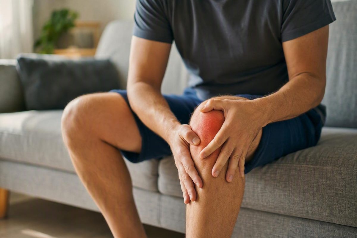
First, decide how urgently the knee needs attention
Important: This article provides general education. It cannot diagnose your knee, determine whether a treatment is safe for you, or replace assessment by a qualified healthcare professional. Knee Pain Zone describes its articles as educational rather than medical treatment in its medical-information notice.
Knee pain and swelling form a symptom pattern, not a diagnosis. The first task is therefore not to identify the exact condition. It is to decide whether you need urgent help, prompt injury assessment, or a brief period of cautious monitoring.
A practical—though not formally validated—triage sequence begins with six questions:
- Was there major trauma? Examples include a fall from height, a forceful direct blow, a vehicle collision, or another high-energy injury.
- Did substantial swelling appear rapidly? Immediate or very rapid swelling after a twist or impact can accompany bleeding or a significant structural injury.
- Does the knee look deformed or misshapen?
- Can you bear weight? Complete inability to put weight through the leg is more concerning than a mild limp.
- Is the joint hot or red, especially with fever, chills, or feeling generally unwell?
- Are there marked calf symptoms? Diffuse calf swelling, redness, or pronounced tenderness changes the urgency.
Seek urgent medical assessment for a hot or red swollen knee accompanied by fever, chills, or malaise. Joint infection is one possible explanation and should not be ruled out at home. Major recent trauma, deformity, complete inability to bear weight, immediate swelling after a twisting injury, and redness or heat with systemic illness are also reasons to seek urgent help (NHS guidance on knee problems).
Arrange prompt injury assessment after major trauma or when you have:
- Visible deformity
- Severe pain
- Complete inability to bear weight
- Immediate substantial swelling after a twisting injury
- A major loss of movement
- Inability to continue the activity at the time of injury
Do not assume that swelling behind the knee or extending into the calf is simply a Baker cyst. Marked calf swelling, redness, or severe tenderness can also occur with a vascular problem that requires immediate evaluation (Hospital for Special Surgery guidance on seeking care).
Certain mechanical symptoms are not necessarily emergencies, but they deserve clinical evaluation rather than repeated cycles of home treatment:
- True locking: The knee becomes physically blocked and cannot move normally.
- Recurrent giving way: The knee repeatedly buckles or feels unable to support you.
- Inability to straighten the knee fully
- Substantial loss of bending or overall motion
Use a lower threshold for contacting the treating team if new swelling develops after a knee replacement, recent knee surgery, or ligament reconstruction. Postoperative advice should come from clinicians who know the procedure, expected recovery, and your individual circumstances.
There is no universal deadline that fits every swollen knee. A mild, improving problem without warning signs is different from recurrent unexplained swelling, rapidly worsening symptoms, or declining function. Urgency depends on the trauma involved, symptom severity and progression, systemic illness, weight-bearing ability, calf symptoms, recurrence, and functional loss.
What knee swelling means—and which details matter
A swollen knee usually means that excess fluid has accumulated inside the joint, around it, or in nearby tissues. Fluid within the joint is commonly called a knee effusion and is sometimes described informally as “water on the knee.” The fluid is not itself a diagnosis: injury, irritation, arthritis, crystals, bleeding, and infection can all be associated with an effusion (Mayo Clinic overview of a swollen knee).
The size difference may be obvious when you compare the knees, but swelling can also feel more prominent than it looks. Accompanying symptoms may include:
- Puffiness around the kneecap or joint line
- A tight or pressurized feeling
- Stiffness
- Warmth or tenderness
- Reduced ability to bend or straighten the knee
- Pain while standing or walking
- Difficulty bearing weight
Where the swelling appears can provide a clue, but not a diagnosis.
- Diffuse swelling: Puffiness involves much of the knee. This can reflect fluid within the joint or more widespread surrounding inflammation.
- Focal swelling: A defined, tender area over the front, inner side, or another specific point may involve a bursa or other local tissue.
- Fullness behind the knee: This can occur with a Baker cyst, but posterior swelling has other possible causes.
- Swelling extending into the calf or leg: This needs particular care when it is pronounced, red, or tender.
Pain location can also help a clinician organize the possibilities. You may be asked whether the pain is at the front, inside, outside, back, or deep within the knee. However, different conditions overlap substantially, so location alone cannot identify the cause. History, examination, and sometimes imaging are needed (Hospital for Special Surgery guide to knee-pain patterns).
The same caution applies to warmth, clicking, popping, grinding, and the movement that triggered the symptoms. These details may help guide an assessment, but none reliably proves that you have arthritis, a meniscus tear, a ligament injury, or an infection.
Before contacting a clinician, record:
- When the pain and swelling began
- Whether an injury occurred
- What movement, impact, or activity preceded it
- How quickly the swelling developed
- Whether the knee feels warmer than the other one
- Whether redness is present
- Whether you can bear weight and walk
- How far the knee can bend and straighten
- Any locking, catching, buckling, or giving way
- Fever, chills, sweats, fatigue, or malaise
- Calf swelling, redness, or tenderness
- Previous knee surgery, replacement, or reconstruction
- Current medicines, particularly anticoagulants
- Similar previous episodes and how they resolved
A short, accurate timeline is usually more useful than choosing a diagnosis from an online symptom list.
Common causes, organized by the pattern of onset
The cause is often narrowed by asking whether symptoms followed one identifiable injury, accumulated with repetitive loading, appeared as a recurrent flare, or began alongside systemic illness. Every pattern below is suggestive, not diagnostic.
| Category | Typical context | Possible accompanying clues | Tests a clinician may consider |
|---|---|---|---|
| Ligament sprain or tear | Twist, awkward landing, sudden change of direction, or force to the knee | Rapid swelling, instability, pain with weight-bearing, or inability to continue the activity | Examination; X-ray when bone injury is possible; sometimes MRI |
| Meniscus injury | Twisting on a planted foot, deep bending, or age-related tissue change | Joint-line pain, later-developing swelling, catching, locking, or limited extension | Examination and, when findings justify it, MRI |
| Cartilage or osteochondral injury | Impact, twist, dislocation event, or repeated joint loading | Deep pain, swelling, catching, or a loose-body sensation | X-ray and sometimes MRI |
| Fracture or dislocation | Fall, direct blow, forceful trauma, or twisting injury | Severe pain, immediate swelling, deformity, or inability to bear weight | X-ray, with further imaging when needed |
| Tendon or bursa irritation | Repetitive kneeling, running, jumping, lifting, or a sudden activity increase | More localized pain or swelling, load-related symptoms, or tenderness over one structure | Clinical examination; ultrasound in selected cases |
| Osteoarthritis | Persistent or recurrent symptoms not linked to one acute event | Stiffness, activity-related pain, reduced motion, or intermittent swelling | Examination and X-rays where appropriate |
| Inflammatory arthritis | Recurrent or persistent inflammatory symptoms, sometimes involving other joints | Warmth, stiffness, swelling, fatigue, or symptoms elsewhere | Examination, imaging, and sometimes aspiration |
| Gout or pseudogout | Sudden inflammatory flare, with or without previous attacks | Marked pain, warmth, redness, and restricted movement | Joint-fluid analysis for crystals; other tests as indicated |
| Bursitis | Repeated pressure, kneeling, impact, or local irritation | Focal swelling over a bursa rather than generalized joint fullness | Examination and sometimes ultrasound or fluid testing |
| Baker cyst | Fullness behind the knee, often alongside another knee disorder | Posterior tightness or discomfort; possible swelling extending downward | Examination and sometimes ultrasound |
| Joint infection | May occur without trauma | Hot or red joint, severe pain, restricted movement, fever, chills, or malaise | Urgent examination and joint-fluid analysis |
These cause-and-clue patterns reflect recognized associations among swelling, injury, overuse, arthritis, crystal disease, bursitis, cysts, and infection; they are not home diagnostic criteria (Mayo Clinic summary of swollen-knee causes).
Acute injury is a broad category, not one diagnosis. A fall, direct blow, awkward landing, or twisting event can affect bone, ligament, meniscus, cartilage, tendon, or several structures at once. Severity cannot be judged solely by whether you heard a pop or remember the precise moment of injury.
Repetitive-load problems may irritate a tendon or bursa, particularly after a sudden increase in running, kneeling, jumping, stair use, lifting, or occupational loading. Activity-related swelling should not automatically be dismissed as harmless overuse. The activity may have aggravated an underlying joint problem rather than being the entire explanation.
Osteoarthritis is one possibility when pain, stiffness, reduced motion, and swelling persist or return without a single acute injury. Symptoms can fluctuate, so variation in swelling from one day to another does not confirm or exclude arthritis.
Inflammatory arthritis and crystal disease can cause striking warmth, redness, tenderness, and swelling. Rheumatoid arthritis, gout, and pseudogout differ in cause and treatment, and redness or warmth cannot distinguish among them. Infection may look similar, which is why a hot or red swollen knee accompanied by systemic illness requires urgent assessment.
Bursitis typically creates more localized swelling over an irritated bursa. By contrast, an effusion arises within the joint and may produce generalized fullness and restricted movement. Appearance alone may not make the distinction clear.
A Baker cyst is a fluid-filled swelling behind the knee and may accompany another disorder within the joint. It describes where fluid has collected, not necessarily why the knee produced excess fluid. A cyst and a vascular problem can produce overlapping posterior-knee or calf symptoms, so attempting to distinguish them by touch or appearance alone is unsafe.
Less common explanations, including bleeding disorders and abnormal tissue growth, may also enter a clinician’s differential diagnosis. These are not the most likely causes for most readers, but persistent unexplained swelling should be assessed rather than self-diagnosed from appearance or location.
What the timing after an injury may suggest
The interval between an injury and visible swelling can help a clinician prioritize the evaluation.
Very rapid or immediate substantial swelling after trauma can suggest bleeding into the joint, known as haemarthrosis. This may accompany a significant ligament injury or a fracture involving the joint. Concern increases when rapid swelling occurs with severe pain, inability to continue an activity, or inability to bear weight.
Swelling that develops later can follow a different pattern. Swelling appearing approximately 24 to 36 hours after injury may reflect a reactive effusion or slower bleeding, including in association with a meniscal injury. Timing, injury mechanism, weight-bearing ability, locking, and systemic symptoms guide the differential diagnosis but do not establish it (clinical review of the acute swollen knee).
A twisting injury can affect a meniscus or cruciate ligament, particularly when the foot is planted and the body rotates. But the mechanism does not identify the damaged structure. Several tissues may be affected by the same movement, and pre-existing joint changes can influence what becomes painful.
Inability to continue an activity immediately after injury raises concern, especially if you also cannot put weight through the leg. That combination may indicate a more severe bone or ligament injury, although an examination and imaging may be needed to determine what happened.
Mechanical symptoms should be described precisely:
- True locking means there is a physical block to normal bending or straightening, not simply that movement is painful.
- Displaced meniscal tissue, a loose body, a cartilage fragment, or another structural obstruction can produce locking.
- Giving way may reflect ligament instability, a mechanical obstruction, pain-related loss of control, or a sensation of instability without a major ligament tear.
Warmth also needs context. A hot or red knee accompanied by fever or malaise raises concern for infection, but warmth alone is not specific. Blood and inflammation after a traumatic injury can also make the knee feel warm as the episode evolves.
Timing and mechanism therefore answer “How quickly should this be evaluated?” more reliably than “Exactly which structure is injured?” Identifying the source requires a physical examination and, when indicated, imaging or joint-fluid analysis.
Safe short-term care for mild symptoms without warning signs
This pathway is only for mild symptoms without:
- Major trauma
- Visible deformity
- Fever, chills, or systemic illness
- A hot, red joint
- Complete inability to bear weight
- Marked calf swelling, redness, or tenderness
- Immediate substantial swelling after a twist
- True locking, recurrent giving way, or inability to straighten the knee
- Recent surgery requiring individualized postoperative instructions
If none of those concerns applies, short-term care can focus on reducing aggravation while preserving tolerable movement.
Use relative rest, not prolonged complete immobilization. Temporarily reduce activities that clearly increase pain or swelling, such as running, deep squatting, kneeling, heavy lifting, or prolonged standing. Continue gentle movement within a comfortable range rather than keeping the knee completely still.
For the first 24 to 48 hours of a new, uncomplicated problem:
- Reduce aggravating activity.
- Elevate the leg while resting.
- Avoid long periods on your feet.
- Gently bend and straighten the knee as symptoms allow.
After this initial period, progressively use the leg more rather than remaining inactive, provided the symptoms are not worsening. Public-health guidance recommends reducing activity initially, preserving gentle movement, and gradually returning to normal use as tolerated (NHS self-care guidance).
Use cold carefully. There is no single ice schedule suitable for every presentation. If cold feels helpful:
- Wrap the cold pack in a cloth or towel.
- Use it for a brief, intermittent session.
- Allow a break between sessions.
- Never place ice directly on the skin.
- Do not use cold where circulation or sensation is impaired.
- Stop if redness, discoloration, blistering, numbness, or pain increases.
Cold can help manage symptoms, but it does not treat every cause of swelling. More icing is not necessarily better.
Compression may help control swelling, but it should feel supportive rather than restrictive. Loosen or remove it if pain increases or the lower leg or foot becomes more swollen. Compression should not be used to postpone assessment of concerning calf symptoms.
Heat may help stiffness in some circumstances, particularly after the early injury period or with a non-acute problem. Stop if it increases swelling or throbbing. Hospital patient guidance supports using a cloth barrier for ice and describes compression as snug rather than a substitute for individualized clinical advice (UMass Memorial guidance on knee pain and swelling).
For stairs, a temporary accessibility strategy can reduce demand:
- Use a handrail when available.
- Lead with the less painful leg going up.
- Lead with the affected leg going down.
- Move one step at a time if needed.
Medication requires an individual safety check rather than a generic dosing recommendation. Acetaminophen may relieve pain, but it is not an anti-inflammatory medicine. Nonsteroidal anti-inflammatory drugs, or NSAIDs, are not suitable for everyone. Kidney disease, stomach ulcers, bleeding risk, anticoagulant use, and interacting medicines can affect whether they are appropriate. Ask a pharmacist or clinician to review your health history and current medicines before advising you (medication safety guidance from the Connecticut Orthopaedic Institute).
Stop home management and seek assessment if:
- Pain or swelling rapidly worsens
- Redness, marked warmth, fever, chills, or malaise develops
- You lose the ability to bear weight
- Marked calf symptoms appear
- Motion or walking deteriorates
- Locking or repeated giving way begins
- Swelling persists without a meaningful improving trend
- The swelling repeatedly returns
Home care should be a monitored trial for a mild presentation—not a way to postpone evaluation while symptoms or function decline.
How clinicians investigate a painful, swollen knee
Evaluation usually begins with a detailed history and physical examination, not with automatically ordering every available scan.
A clinician may ask:
- Was there trauma, twisting, or direct impact?
- How quickly did the swelling appear?
- Has this happened before?
- Where is the pain?
- Can you bear weight?
- Does the knee lock, catch, or give way?
- Can you bend and straighten it?
- Is there fever, warmth, redness, or systemic illness?
- Does pain occur at night?
- Have you had knee surgery or a joint replacement?
- Which medicines do you take, including anticoagulants?
- Do other joints hurt or swell?
During the examination, the clinician may compare both knees, determine whether swelling is diffuse or focal, locate tenderness, assess motion, observe walking or another functional task, and check stability when it is safe to do so. The aim is to decide which broad category is most plausible and what question—if any—testing needs to answer.
Testing is question-driven:
- X-rays can identify fractures or dislocations and show evidence consistent with arthritis.
- Ultrasound may help assess selected tendon, ligament, fluid, or cyst concerns.
- MRI can reveal deeper soft-tissue injuries that are not visible on an X-ray.
- Joint aspiration, also called arthrocentesis, may be used when examining the fluid can help clarify the cause.
During aspiration, a clinician removes joint fluid with a needle. The sample can be checked for blood, bacteria, and crystals associated with gout or pseudogout. Removing fluid may also reduce pressure, pain, or stiffness, but it does not necessarily treat the underlying problem or prevent fluid from returning (Mayo Clinic guide to swollen-knee diagnosis and treatment).
The examination may extend beyond the knee. Hip or spinal problems can sometimes refer pain toward the knee, so a clinician may assess those areas when the symptom pattern or knee findings do not fully explain the pain.
Not every person needs aspiration, and not every painful knee needs an MRI. A normal X-ray does not exclude all soft-tissue problems, while a finding on MRI does not automatically prove that it is responsible for the symptoms. Test results need to be interpreted alongside the history and examination.
Treatment depends on the cause, not swelling alone
Treatment can be organized around four purposes:
- Address the underlying cause
- Control pain, pressure, and swelling
- Restore motion, strength, balance, and confidence
- Prevent recurrent loss of function
The balance among these aims differs by diagnosis. An infection, fracture, inflammatory flare, tendon problem, ligament sprain, and osteoarthritis-related effusion should not receive one interchangeable plan.
Rehabilitation may include:
- Range-of-motion work
- Progressive strengthening
- Balance or control exercises
- Walking or movement retraining
- Temporary activity modification
- Gradual progression back to normal demands
Physical therapy may be considered for selected meniscus injuries, ligament sprains, osteoarthritis, patellofemoral pain, and rehabilitation after a fracture. The program should reflect the diagnosis, current function, symptom irritability, and activity goals rather than a universal exercise list.
A brace may provide support or limit a particular movement in some circumstances. It is not a treatment for every swollen knee, and it should not create false reassurance about an undiagnosed injury.
Aspiration can serve two purposes. Diagnostically, the fluid may reveal blood, bacteria, or crystals. Symptomatically, removing fluid may temporarily reduce pressure and stiffness. If the underlying driver remains active, the fluid can return.
Anti-inflammatory medication may be appropriate for selected inflammatory or musculoskeletal problems after reviewing medical history, current medicines, and relevant risks. A corticosteroid injection may also be considered in particular circumstances, but it is not an automatic treatment for unexplained swelling. The possible cause of the swelling must be considered first.
Arthroscopy or another operation may be considered for selected structural injuries, repairable tissue damage, loose material within the joint, instability, fracture-related problems, or severe joint disease. Swelling alone is not an indication for surgery. Management of an acutely swollen knee is cause-dependent and can range from medication and rehabilitation to surgery for appropriate structural abnormalities (clinical review of swollen-knee diagnosis and management).
It helps to separate symptom relief from treatment of the cause:
- Ice may reduce discomfort but does not repair a ligament or treat infection.
- Compression may limit swelling but does not identify why fluid accumulated.
- Pain medicine may make movement easier without resolving the underlying disorder.
- A brace may improve support without correcting every source of instability.
- Drainage may relieve pressure without preventing recurrence.
No exercise program, injection, brace, medicine, drainage procedure, or operation is the best option for every painful, swollen knee. The appropriate choice follows the diagnosis.
Monitoring recovery and returning to normal activity
Recovery is better judged by direction and function than by a calendar alone. A mild overuse flare, ligament injury, crystal flare, and persistent arthritis-related effusion will not follow the same timeline.
Signs of a meaningful improving trend include:
- Swelling is settling rather than expanding.
- The knee bends and straightens more easily.
- Walking is becoming smoother and more stable.
- Ordinary daily activities are better tolerated.
- The knee is not repeatedly locking or giving way.
- Activity does not produce a substantial or lasting flare.
Progress gradually. Begin with gentle motion and routine daily tasks, then increase walking and other demands as tolerated. More demanding activity can follow when swelling is minimal or continuing to settle, motion is improving, walking is stable, and the proposed activity does not trigger a meaningful increase in pain, swelling, or instability.
Postpone sport and heavy lifting while discomfort remains significant or movement is poor. Being able to force your way through an activity is not the same as tolerating it well. A substantial flare in swelling or function is a signal to reduce the load and reconsider the progression.
Rehabilitation commonly uses gradual activity progression alongside strengthening, movement modification, balance work, and range-of-motion exercises, but the plan should be adapted to the condition and the person (Hospital for Special Surgery overview of physical therapy for knee pain).
Arrange professional assessment if symptoms:
- Worsen instead of improving
- Do not show a meaningful improving trend
- Repeatedly return
- Interfere with work, sleep, walking, or daily activities
- Require ongoing self-treatment just to maintain basic function
Recurrent locking, giving way, inability to straighten the knee, or recurrent effusion should not be treated as a conditioning problem alone. These patterns may require examination for mechanical, inflammatory, or other underlying causes.
Before the appointment, prepare:
- A short symptom timeline
- The injury mechanism, if any
- How quickly swelling appeared
- Photos showing changes in swelling, when useful
- Your current ability to walk and use stairs
- Details of locking, catching, or instability
- Fever, warmth, redness, or calf symptoms
- Previous episodes and treatments
- Prior operations or joint replacement
- A complete medicine and supplement list
- The activities you need or hope to return to
Frequently asked questions
Is a hot, red, swollen knee an emergency?
It can be. A hot or red swollen knee accompanied by fever, chills, sweats, malaise, or rapidly worsening illness requires urgent medical assessment because infection is possible. Redness and fever can accompany a knee infection, and rapidly developing extreme swelling may require urgent intervention (Hartford HealthCare guidance on knee swelling).
Warmth without systemic illness is less specific because bleeding or inflammation after an injury can also make the knee feel warm. Even so, marked redness, increasing pain, unexplained swelling, or worsening illness should lower the threshold for urgent assessment.
Can I walk on a swollen knee?
It depends on the severity and cause. With mild symptoms and no warning signs, short periods of comfortable walking and gentle movement may be reasonable. Reduce the distance or load if walking meaningfully increases pain, swelling, or limping.
Do not force weight-bearing after major trauma, with visible deformity, or when you cannot put any weight through the leg. Complete inability to bear weight warrants prompt assessment. Repeated buckling or a physically blocked knee also needs evaluation rather than an attempt to “walk it off” (Connecticut Orthopaedic Institute guidance on home care and assessment).
Should I use ice or heat for knee pain and swelling?
For a new, uncomplicated problem, protected intermittent ice may help manage symptoms. Place a cloth between the skin and cold source, use brief sessions with breaks, and never apply ice directly to the skin.
Heat may help stiffness in some circumstances, especially after the early injury period, but stop if swelling or throbbing increases. Avoid heat or ice where circulation or sensation is impaired, and stop if you notice increasing redness, discoloration, blistering, or worsening symptoms.
Neither ice nor heat identifies or treats every cause. A hot or red knee with systemic illness needs medical assessment, not a temperature-based home remedy.
What does swelling behind the knee mean?
Fullness behind the knee may be a Baker cyst, which can occur alongside another disorder within the joint. It may produce tightness or discomfort and can sometimes extend toward the calf.
Posterior swelling has other possible causes, however, and symptoms alone cannot safely distinguish a cyst from a vascular problem. Seek prompt assessment for marked calf swelling, redness, significant tenderness, or rapidly worsening symptoms.
What can fluid drained from the knee reveal?
Joint fluid can be examined for blood, which may follow injury or a bleeding problem; bacteria, which may indicate infection; and crystals associated with gout or pseudogout (Mayo Clinic explanation of joint aspiration).
Aspiration may also reduce pressure and stiffness temporarily. It does not necessarily cure the cause, and fluid can return if the condition producing it remains active. The result is interpreted together with the history, examination, and any imaging or other tests.
The practical next step: Seek urgent help for infection signs, major trauma, deformity, complete inability to bear weight, rapid substantial swelling, or concerning calf symptoms. Use brief, cautious self-care only for mild symptoms without warning signs. Arrange assessment when swelling worsens, recurs, limits function, or does not show a meaningful improving trend. The pattern supplies clues, but an accurate diagnosis determines the appropriate treatment.